![]()

G.D. Kotzalidis, A. Facchi, L. Tarsitani, V. Mantua, P.P. Colombo, P. Pancheri - Vol. 7, Giugno 2001, num.2
Testo Immagini Bibliografia Summary Riassunto Indice
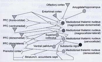
Thalamic abnormalities in schizophrenia
Anomalie talamiche nella schizofrenia
Tab. I. Thalamic volume in schizophrenia: structural studies in vivo. Volume talamico nella schizofrenia: studi strutturali in vivo.
|
Paper |
Technique |
Sample |
Results |
|
Portas et al., 1998 (29) |
MRI |
15 patients vs. |
No significant difference, but correlation between reduced volume on one side and prefrontal cortex and thalamocortical projection abnormalities as well as positive symptoms in schizoprenics on the other |
|
Wolkin et al., 1998 (30) |
MRI T1-weighted |
25 schizophrenic patients |
No differences between schizophrenics and controls in thalamic size or parathalamic white matter, but signal hypointensity was found in schizophrenic periventricular (cerebellar vermis, occipital tail, peri-callosal, peri-caudate and peri-fornical areas) and cortical structures (frontal, temporal, posterior parietal cortex, middle occipital gyrus and prefrontal cortex, specially at left) |
|
Staal et al., 1998 (31) |
MRI T1-and T2-weighted |
32 same-sex siblings |
Thalamic volume was smallest in schizophrenic patients and smaller with respect to their healthy siblings, who also had smaller volumes compared to that of normal controls |
|
Arciniegas et al., 1999 (32) |
MRI T1-weighted |
10 men and 11 women with schizophrenia, paranoid subtype, vs.15 healthy men and 12 healthy women |
No difference between schizophrenic patients and healthy controls in thalamic volume and no effect of gender, diagnosis or hemispheric laterality |
|
Dasari et al., 1999 (33) |
MRI T2-weighted |
20 schizophrenic adolescents |
No differences between schizophrenic and bipolar adolescents in thalamic volume, but significant volume reduction in both groups as compared to healthy controls |
|
Lawrie et al., 1999 (34) |
MRI T2-weighted |
10 first-episode schizophrenics vs. |
Relatives of schizophrenic patients have smaller thalami bilaterally, both with respect to schizophrenics and to controls; schizophrenics have reduced volume of the amygdala/hippocampal complex with respect to both relatives and controls |
|
Seidman et al., 1999 (35) |
MRI T1-weighted |
28 non-psychotic/ non-schizotypal first degree relatives of schizophrenics
|
Significantly reduced volume of thalamus and amygdalar/hippocampal complex, marginal reduction in pallidum, striatum, cerebellum in relatives of schizophrenic patients as compared to normal controls |
|
Andreasen et al., 1994 (6) |
Averaged MRI |
39 schizophrenic men |
Smaller thalami in schizophrenic patients, more in lateral than in medial nuclei, decreased cortical size mostly in frontal, less in parietal and temporal cortex |
Tab. II. Post-mortem studies of thalamus in schizophrenia. Studi post mortem del talamo nella schizofrenia.
|
Paper |
Technique |
Sample |
Results |
|
Popken et al., 2000 (61) |
Post-mortem thionin- Nissl staining, in situ hybridisation; stereological cell and volume estimates |
6 schizophrenic patients vs. 6 matched controls for age, sex and post-mortem interval (autolysis) |
Decreased neuronal numbers in the parvocellular and densocellular portions of the mediodorsal thalamic nucleus, but not in total volumes of thalamic nucleior in neuronal numbers of the magnocellular portion of the mediodorsal thalamic nucleus or the ventral posterior medial thalamic nucleus |
|
Falke et al., 2000 (62) |
Post-mortem immunohisto-chemistry numbers, and stereological counting |
11 elderly schizophrenic patients vs. 11 elderly healthy controls vs. 12 patients with Alzheimers disease |
No differences between schizophrenics and controls in thalamic or striatal neuronal neurofibrillary tangles, astrocytosis or microgliosis |
|
Young et al., 2000 (63) |
Post-mortem optical dissection for obtaining stereological cell and volume estimates; Nissl-cresyl violet staining |
8 schizophrenic men vs. 8 age-matched healthy men |
Reduced neuronal numbers (-35%) and volume (-24%) in the mediodorsal thalamic nucleus and anteroventral/anteromedial thalamic nucleus neuronal number (-16%) in schizophrenic patients, as compared to controls |
|
Pakkenberg, 1992 (64) |
Post-mortem stereometry (unbiased Cavalieri volume estimator); staining |
8 drug-naïve schizophrenic patients vs. 8 controls; 12 neuroleptic-treated schizophrenics vs. 11 controls |
Reduced volume of the mediodorsal thalamic nucleus in drug naïve (-31%) and neuroleptic- treated (-22%) schizophrenic patients (but haematoxylin/eosin fixation time differed between the two groups, thus rendering shrinkage a possible explanation) |
|
Danos et al., 1998 (65) |
Post-mortem parvalbumin-immunocytochemistry plus Nissl-myelin staining |
12 schizophrenic patients vs. 14 controls |
Schizophrenic patients have significantly reduced densities of parvalbumin-positive thalamocortical projection neurones (glutamatergic) in right and left anteroventral thalamic nuclei and non-significantly reduced densities of all anteroventral thalamic neurones |
|
Davidsson et al., 1999 (66) |
Post-mortem Western blotting, rab.3a and synaptophysin immunochemistry |
19 schizophrenic patients vs. 39 age- matched controls |
Decreased rab.3a in the thalamus, frontal and parietal cortex of schizophrenic patients as compared to controls, decreased rab.3a and synaptophysin in the gyrus cinguli and the hippocampus in schizophrenics and no difference between schizophrenics and controls for both proteins in temporal cortex or cerebellum |
| Blennow et al., 2000 (67) | Post-mortem Western blotting, rab.3a immunochemistry | 22 schizophrenic patients vs. 24 controls not differing for age | Decreased rab.3a in schizophrenic left and right (less) thalamus, hippocampus, cingulate, frontal and parietal cortex; no difference in temporal cortex and cerebellum |
| Blennow et al., 1996 (68) | Post-mortem Western blotting, rab.3a immunochemistry | 19 schizophrenic patients vs. 39 age- matched controls | Decreased rab.3a in schizophrenic patients, which is more pronounced at the left thalamus and suggests decreased synaptic density resulting in disconnection with the corticolimbic pathways, hence giving place to psychotic symptoms. Rab.3 is a protein associated with both synapses and vesicles |
| Landén et al., 1999 (69) | Post-mortem radioimmunoassay for synaptophysin and chromogranins thalamus | 9 schizophrenic patients vs. 9 age-matched controls | Reduced synaptophysin in the left thalamus of patients with schizophrenia compared to controls; but no difference in left or right thalamic chromogranins or synaptophysin in the right |
| Court et al., 1999 (70) | Post-mortem autoradiography of nicotine receptor sub- types (cytisine and alpha bungarotoxin) | 12 schizophrenic patients vs. 12 age- matched controls vs. 14 patients with Lewy body dementia | Moderate alpha-bungarotoxin binding reduction in the reticular thalamic nucleus in schizophrenics vs. controls and strong reduction in patients with dementia with Lewy bodies; nicotine binding reduced only in Lewy body dementia in the lateral dorsal nucleus of the thalamus |
|
Richardson-Burns et al., 2000 (71) |
Post-mortem in situ hybridisation to assess expression of mGluR1, 2, 3, 4, 5, 7 and 8 in various thalamic nuclei | 12 elderly schizophrenic patients (6 drug-free at the time of death) vs. controls not differing from patients in age, post- mortem interval or cause of death | No differences in thalamic metabotropic glutamate receptor expression between schizophrenics and controls in anterior, dorsomedial, lateral dorsal, central medial, reticular, and nuclei of the ventral tier |
|
Ibrahim et al., 2000 (72) |
Post-mortem in situ hybridisation for ionotropic glutamate autoradiography of the same receptors | 12 DSM-III-R schizophrenics vs. 8 non-psychiatric | Decreased NMDAR1 (mediodorsal and centromedial nuclei), NMDAR2B (centromedial n.) and C (anterior, mediodorsal, laterodorsal receptor mRNA; controls and centromedial nuclei), gluR1 (mediodorsal and centromedial nuclei) and 3 (centromedial nucleus), and KA2 (anterior, mediodorsal, laterodorsal, centromedial and ventral thalamic nuclei) mRNA; decreased polyamine (anterior, mediodorsal and centromedial nuclei) and glycine (anterior nucleus) binding sites of the NMDA receptor |
Tab. III. Functional imaging studies of the thalamus in schizophrenia. Studi di visualizzazione cerebrale funzionale del talamo nella schizofrenia.
|
Paper |
Technique |
Sample |
Results |
|
Omori et al., 2000 (73) |
Proton magnetic resonance spectroscopy |
20 schizophrenic patients vs. 16 age- matched healthy controls |
Lower N-acetyl aspartate/total creatine derivatives and choline-derived compounds/total creatine derivatives in schizophrenic patients as compared to controls in the thalamus, but not in the frontal cortex |
|
Deicken et al., 2000 (74) |
Proton magnetic resonance spectroscopy |
17 medicated schizophrenic men vs. 10 age-matched healthy male controls |
Lower N-acetyl aspartate/total creatine derivatives but not choline-derived compounds/total creatine derivatives in schizophrenic patients as compared to controls in the thalamus, bilaterally |
|
Rodríguez et al., 1997 (75) |
99Tc-labelled hexamethyl- propylene- aminoxime(HMPAO) and SPECT |
39 schizophrenics tested while on classical neuroleptics and after 6 months on clozapine vs. healthy subjects (database) |
Lower thalamic, right prefrontal cortical and striatal perfusion with respect to control values in neuroleptic non-responders; responders to clozapine had their perfusion normalised in the same areas |
|
Hazlett et al., 1999 (76) |
Co-registered MRI and PET [18F]Fluorode- oxyglucose (FDG) under serial verbal learning task |
27 schizophrenic patients vs.13 patients with schizotypal personality disorder vs. 32 healthy controls |
No significant differences in total thalamic volume among the three groups, but schizophrenic patients showed decreased metabolism in the medio-dorsal thalamic nucleus bilaterally and less pixels in the left anterior thalamic region, while schizotypal personality patients had less pixels in the right medio-dorsal nucleus with respect to controls |
|
Buchsbaum et al., 1996 (77) |
[18F]fluoro- deoxyglucose PET and MRI |
20 drug naïve schizophrenic patients vs. 15 healthy controls |
Reduced metabolic rate in right thalamus in schizophrenic patients with respect to controls and loss of the normal asymmetry (right > left); smaller thalamic volume in schizophrenics, particularly in the left anterior region |
|
Andreasen et al., 1996 (13) |
MRI on [15O]water PET rCBF and practised and novel recall tasks from Wechsler Memory Scale |
14 schizophrenic patients (11 drug-free [> 3 weeks] and 3 drug-naïve; 10 men and 4 women) vs. 13 healthy controls (6 men and 7 women) |
Decreased regional blood flow in patients, as compared to controls, with performance of practised task in right medial and lateral frontal cortex, left thalamus and left cerebellum; with the novel task, these same regions show even more important reductions in patients, as well as reductions also in the anterior cingulate gyrus, in the mammillary bodies, bilaterally in the temporal cortex and in the lenticular nuclei |
|
Crespo-Facorro et al., 1999 (11) |
MRI on [15O]water PET rCBF and practised and novel recall tasks from Rey Auditory Verbal Learning Test |
14 schizophrenic patients (11 drug-free [ > 3 weeks] and 3 drug-naïve; 10 men and 4 women) vs. 13 healthy controls (6 men and 7 women) |
Decreased regional blood flow in patients, as compared to controls, with performance of practised task in left dorsolateral prefrontal cortex, bilateral medial frontal cortex, left supplementary motor area, left thalamus, left cerebellar regions, anterior vermis, and right cuneus; with the novel task, reductions in patients, as compared with controls, were observed in the right anterior cingulate, right thalamus, and bilateral cerebellum (left greater than right) |
|
Siegel et al., 1993 (78) |
[18F]fluoro- deoxyglucose PET |
70 drug-free ( > 4 weeks) schizophrenic men vs. 30 healthy age-matched men |
Schizophrenic patients showed decreased metabolism in medial frontal cortex, cingulate gyrus, medial temporal lobe, corpus callosum and ventral caudate and increased metabolism in left lateral temporal cortex and occipital cortex with respect to controls; inverse correlation between medial frontocortical and thalamic activity and total, positive and negative BPRS scores |
|
Kim et al., 2000 (79) |
[15O]H2O PET rCBF while subjects stay with eyes closed and no instruction as to mental activity (resting state) |
30 chronic, neuroleptic-free ( > 3 weeks) schizophrenic patients vs. 30 healthy controls |
Lower regional blood flow in prefrontal cortex and higher in thalamus and cerebellum in schizophrenic patients, as compared to controls; cerebral blood flow alterations in chronic schizophrenic patients are much similar to those occurring in acute episode patients |
|
Andreasen et al., 1997 (10) |
[15O]H2O PET rCBF while subjects stay with eyes closed and no instruction as to mental activity (resting state) |
17 neuroleptic naïve first-episode schizophrenic patients vs. healthy controls |
Schizophrenic patients display decreased perfusion with respect to controls in the lateral, medial and orbital prefrontal cortex, inferior temporal and parietal cortices, whereas increased perfusion was found in schizophrenics in the thalamus, the cerebellum, and the retrosplenial cingulate |
|
OLeary et al., 1996 (9) |
[15O]H2O PET rCBF while subjects perform an attentional task |
10 drug-free/naïve schizophrenic patients vs. 10 healthy controls |
Schizophrenic patients do not activate the right superior temporal gyrus as much as controls, but have higher regional cerebral blood flow in the left superior temporal gyrus during activation; these findings point at a deficit in thalamocortical integration |